Retinal imaging allows us to see the inside of your eye without dilation. We use Optomap® to get a detailed, wide-angle image of the retina, so we can catch early signs of eye disease and track changes.
Retinal Imaging with Optomap® in Maple Glen, PA
A Wider View of Your Retinal Health
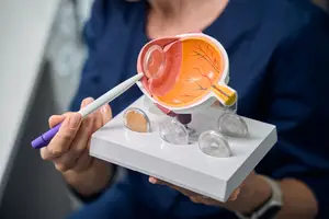
Optomap captures a high-resolution image of the retina, the light-sensitive tissue at the back of your eye. Unlike traditional methods that view only a narrow portion of the eye, Optomap shows up to 200 degrees in a single image, helping us see areas that standard scopes can miss. It’s quick, non-invasive, and suitable for most patients.
Why We Use Retinal Imaging
Many serious eye conditions develop without symptoms.
Optomap helps us detect early signs of problems like retinal detachments, glaucoma, macular degeneration, diabetic retinopathy, and hypertension-related changes, often before you notice vision changes.
It also gives us a clear baseline to compare from year to year.

No Dilation Needed for Most Patients
In many cases, Optomap lets us skip dilation drops entirely.
That means less light sensitivity, less blurriness, and a quicker visit, especially helpful if you’re driving yourself or need to return to work right after your appointment.
In some cases, dilation may still be recommended based on your eye health history.
Retinal Imaging as Part of Routine Care
We offer Optomap imaging during most annual comprehensive eye exams. It’s a useful tool for preventive eye care and for monitoring ongoing eye conditions.
If you’re managing diabetes, high blood pressure, or any chronic eye condition, retinal imaging helps us keep track of subtle changes that might need attention.

What to Expect During Your Imaging
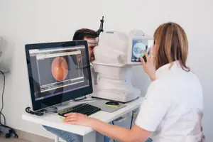
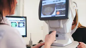
The process is simple: you sit at the machine, look into the lens, and see a small flash of light as the image is captured. It only takes a few seconds per eye.
Your eye doctor will then review the results with you, show you the image, and explain anything we see.
Ask About Optomap at Your Next Visit
If you’re due for a comprehensive eye exam or want to better understand your retinal health, let us know. At Maple Glen Eyecare, our team will explain if Optomap is right for you and make sure you feel comfortable every step of the way.